Hungarian Innovation Breakthrough: Electrical Activity of Neurons and Their Processes in the Continuously Moving Brain Becomes Measurable for the First Time During Brain Surgery

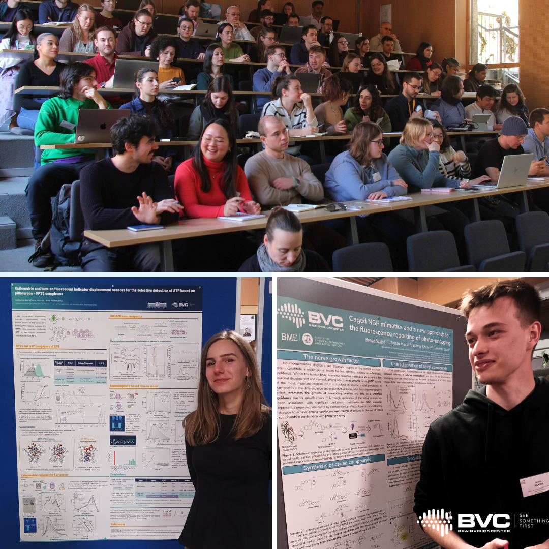
A scientific paper by Dr. Balázs Rózsa (BrainVisionCenter, Institute of Experimental Medicine), Dr. Attila Losonczy (UT Southwestern Medical Center), and their co-authors was published in the March issue of Neuron, one of the world’s leading journals in neuroscience. The study, titled “Movement-stabilized three-dimensional optical recordings of membrane potential changes and calcium dynamics in hippocampal CA1 dendrites,” presents a three-dimensional real-time motion-correction method developed through the collaboration of BrainVisionCenter (BVC)—founded by Botond Roska and Balázs Rózsa—and Femtonics Ltd.

Figure 1. Real-time motion correction and single-cell dual-color labeling for motion-stabilized dendritic voltage-calcium imaging in vivo

This technology has, for the first time, made it possible to measure the brain’s elementary functional units—neurons and their processes, the so-called dendrites—in a continuously pulsating, moving brain, at the extremely fast timescales characteristic of brain activity, reaching speeds of several kilohertz. Under the leadership of Attila Losonczy and Balázs Rózsa, the researchers implemented rapid image stabilization together with simultaneous high-resolution, AI-assisted measurements in three dimensions.

The instrument—protected by more than 50 international patents—is manufactured and distributed by Femtonics Ltd. Its users include leading universities and research institutes such as Stanford University, Yale University, Columbia University, Harvard University, Boston University, the University of Oxford, McGill University, the University of Helsinki, MIT, and Caltech.

The publication in Neuron marks the 25th high-impact paper published in a Nature Index journal generated by the Institute of Experimental Medicine and BrainVisionCenter using a Femtonics instrument. This is considered a record achievement in the Hungarian high-tech sector. Globally, several hundred high-ranking scientific publications have been published using Femtonics laser microscopes. Revenue generated from the sale of this instrument platform has exceeded HUF 10 billion, which the company has reinvested in full into medical research and innovation.

The technology presented in the Neuron paper could enable intraoperative measurement of activity in the pulsating human brain, opening new avenues in medical diagnostics and therapy. It is capable of tracking motion of up to 1 mm while measuring the moving brain with 100-nanometer precision—approximately one-thousandth of the thickness of a human hair. Researchers at BVC and Femtonics are now working on the development of suitable sensors for human brain measurements, paving the way toward clinical application.

The key scientific breakthrough enabled by this invention is that dendritic integration and back-propagating action potentials—previously studied mainly in vitro—can now also be observed in awake, behaving animals, and tracked along the complex, tree-like branching processes of neurons, while filtering out motion-related artifacts arising from brain tissue movement, respiration, and heartbeat. The system can reposition the measurement coordinate system in less than 100 microseconds (i.e. 0.0001 seconds), thereby compensating for tissue displacement. In essence, it functions as a high-speed 3D image stabilizer used during laser scanning, resulting in stable images and signals free from motion noise.

One of the method’s major innovations is that, instead of using previously introduced artificial fluorescent beads in the brain, the neuronal soma itself can serve as the reference point for motion tracking. Because this approach does not trigger an immune response, it avoids damage to the brain and enables more accurate measurements. In addition, the method reduces motion-induced measurement artifacts by more than two orders of magnitude.

Figure 2. Electrical coupling properties between soma and dendrites across the dendritic arbor of hippocampal CA1 pyramidal neurons

Imaging based on the widely used calcium sensors provides only indirect information about electrical activity, and therefore offers a more temporally blurred view of brain function. By contrast, in the present study conducted by UT Southwestern Medical Center, BrainVisionCenter, and the Institute of Experimental Medicine, newly developed genetically encoded voltage sensors, used alongside calcium sensors, directly report electrical changes in the brain with a temporal precision of up to one ten-thousandth of a second. Until now, such measurements had not been feasible in vivo, at subcellular resolution and in 3D, because no sufficiently fast measurement technology existed.

The required technological breakthrough was provided by Femtonics’ acousto-optic technology, which enabled 3D measurements that are 6–7 orders of magnitude faster. This dual functional labeling approach allowed researchers to visualize electrical signals and the calcium responses they evoke at the same time. For labeling, Attila Losonczy’s group developed a novel rapid genetic delivery method based on in vivo electroporation, which yielded stronger contrast and better functional signals than any previous method, while leaving the brain unharmed.

The results show that responses measured in individual dendritic spines become progressively weaker as signals back-propagate along neuronal processes, the so-called dendrites. In contrast, trains of action potentials generate more sustained depolarization, allowing information to reach distal dendrites more reliably. Signals evoked by targeted photostimulation showed the same distance-dependent attenuation, suggesting that dendritic filtering arises primarily from the neurons’ own intrinsic properties. In addition, the researchers detected local dendritic voltage events that were independent of activity in the neuronal soma. This indicates that individual dendritic branches may function as partially autonomous computational subunits, almost like independent processors.

Another important finding is that—contrary to the currently prevailing view—the relationship between voltage signals and calcium signals weakens progressively as the number of neuronal branch points increases. In distal dendritic compartments, voltage signals remain measurable, but the corresponding calcium responses decline more rapidly, especially beyond branch points. This suggests that the structure and electrical properties of dendrites influence not only the propagation of voltage, but also calcium influx, thereby enabling branch-specific plasticity and molecular function.

These findings bridge the gap between in vitro dendritic measurements and in vivo neuronal dynamics. They establish a technological foundation for the direct study of dendritic computation during learning and memory formation in awake, behaving animals.

The article is available at:
https://www.cell.com/neuron/abstract/S0896-6273(26)00004-8

Our Young Researchers Represented Hungary at an International Scientific Seminar in France

Kiss Etelka and Szabó Bence, PhD researchers from our institute, recently participated in a prestigious one-week scientific seminar held at the École de Physique des Houches in France. Notably, they were the only attendees representing Hungary. 

The event brought together 50 young researchers from around the world, who engaged in discussions across multiple courses focusing on scientific innovations at the intersection of physics, chemistry, and biology. Lectures delivered by internationally renowned experts aimed not only to share cutting-edge knowledge but also to empower participants to implement these insights within their own laboratories. 

Beyond the scientific program, the seminar offered valuable opportunities for international exposure and professional networking. 

We are especially proud that our colleagues had the opportunity to present their ongoing research to a distinguished scientific audience. Their work focuses on the development of labeling compounds used in microscopy, as well as the associated methods and procedures, contributing to advancements in biological research. 

Two of Our Young Researchers Earn PhD Degrees with “Summa Cum Laude” Distinction

We are proud to announce that two of our young researcher colleagues, Attila Csomos and Farkas Domahidy, have recently obtained their PhD degrees. It is a particular pleasure that both were awarded the “summa cum laude” distinction by the Scientific Committee, reflecting their outstanding scientific achievements. 

Attila Csomos conducts research in the Chemical Development team at BrainVisionCenter, focusing on small-molecule fluorophores and functional probes for microscopy. His doctoral work explored the conformational locking of the chromophore of the Green Fluorescent Protein (GFP), with the aim of enabling its application in small-molecule form for fluorescence imaging. His research resulted in the development of a novel family of fluorescent zinc sensors, as well as two new heteroaromatic fluorophore scaffolds. These advances open new possibilities in neuroscience research by providing stronger fluorescence signals, particularly when used with the ATLAS two-photon microscopes developed at BVC. 

Farkas Domahidy, a chemical engineer, focuses his research on DNA-binding fluorescent probes. His PhD work investigated the relationship between molecular structure and fluorescence behavior, with particular emphasis on thiazole orange derivatives. A key outcome of his research is a comprehensive study that, for the first time, systematically compares the fluorescence enhancement and PCR applicability of the most widely used thiazole-orange-based DNA-binding dyes. This work enables a more informed selection of dye compounds for PCR-based applications. In addition, his research includes the development of new DNA-binding dye molecules and the investigation of their fluorescence enhancement mechanisms. 

The achievements of our colleagues highlight not only their individual excellence but also the strength of BrainVisionCenter’s interdisciplinary research environment, where chemistry, biology, and advanced imaging technologies come together to drive new scientific discoveries. 

We warmly congratulate Attila and Farkas on their outstanding accomplishments and wish them continued success in their scientific careers. 

BrainVisionCenter at Brain Awareness Week at ELTE

Researchers at the BrainVisionCenter participated in the Brain Awareness Week event series hosted at Eötvös Loránd University (ELTE), where they showcased their work to hundreds of interested students and offered a glimpse into the world of neuroscience. 

Our researchers are no strangers to education or to introducing the next generation to the mysteries of scientific life-especially when they can do so through engaging and interactive demonstrations. During the event, visitors explored key concepts of brain research using a LEGO mouse model, alongside our world-first device, Moculus. 

The Moculus is the result of a collaboration between the BrainVisionCenter and the HUN-REN Institute of Experimental Medicine (HUN-REN KOKI). This virtual reality (VR) headset, specifically optimized for mice, opens up entirely new perspectives in the study of brain function, particularly in visual and sensory processing. 

By using Moculus, our researchers contribute to advances in restoring vision, as well as to improving the diagnosis and future treatment of central nervous system disorders. 

The event provided an excellent opportunity to bring science closer to the younger generation and to demonstrate how curiosity can evolve into a career in research. 


Young Talents at Our Research Institute

It is always a special pleasure for us to welcome young, talented students to our Research Institute and give them a glimpse into the world of science.  

Recently, students from Eötvös High School in Budapest visited us; they were given this opportunity in recognition of their outstanding results in the János Irinyi National Chemistry Competition. During the visit, they were able to experience firsthand how research work is conducted in practice and the environment in which the latest scientific findings are generated.  

The program not only provided a glimpse into everyday laboratory life but also served as a source of inspiration for the participants:  

“It was fascinating to see a functioning lab, especially one dedicated to brain research. Thanks to this, I can better imagine what a researcher’s daily life looks like.”  

“It was great to visit such a high-level research facility, get a glimpse of the working conditions, and see how well-equipped a laboratory in Hungary can be. I would have happily spent more time there.”  

“Overall, this program inspired me to consider pursuing research in chemistry in the future. I would definitely recommend this opportunity to anyone who asks!” 

We believe that opportunities like these help bring the next generation even closer to science-and perhaps the researchers of the future are being born right now. 

BrainVisionCenter researcher gives lecture to high school students about neuroscience

Balázs Tarján, biologist researcher at our institute, recently gave a lecture at Óbuda University for science-oriented secondary school students from Budapest and Hungarian communities beyond the border.
In his talk titled “What Does a Neuroscientist Do?”, he introduced the everyday work of a researcher – from conducting animal experiments and reading and writing scientific articles to giving lectures and preparing research grant proposals.
The students showed great interest and asked several questions, particularly about the research conducted at BrainVisionCenter aimed at restoring vision.

Balázs Rózsa was a guest of the American Chamber of Commerce’s Healthcare Committee

It was a great honor for us that the American Chamber of Commerce (AmCham)’s Healthcare Committee invited our company’s Scientific Director, Balázs Rózsa, to a professional discussion.

The committee members, who represent leading figures in the pharmaceutical industry, the medical technology sector, and private healthcare providers, listened to his presentation with great interest. During the discussion, Balázs provided insight into his professional career, his personal motivations, and the background and vision behind the founding of BrainVisionCenter.

The event showcased BrainVisionCenter’s pioneering technological solutions and equipment, which are opening up new horizons in neuroscience research and therapeutic development. Particular emphasis was placed on the areas of application where our work is already having a significant impact, including:

  • vision restoration
  • epilepsy treatment
  • Alzheimer’s disease research

During the professional discussion that followed the presentation, the potential for collaboration in pharmaceutical research was also discussed. The participants’ active interest clearly demonstrated the growing demand for innovative, multidisciplinary approaches capable of accelerating translational research. The significance of interdisciplinary research was a key theme in the responses to the questions. The projects underway at the BrainVisionCenter are based on the close integration of medicine, neurophotonics, artificial intelligence, and advanced technological solutions, enabling the development of new diagnostic and therapeutic approaches.

Thank you for the invitation and the opportunity for this valuable professional exchange.

We’ve joined the European Animal Research Association (EARA)

We are pleased to announce that BVC has become the fourth organization in Hungary to join the European Animal Research Association (EARA), an international professional community, represents credible, evidence-based, and transparent communication working with animals in scientific research across Europe.

EARA brings together more than 200 member organizations and actively supports responsible professional dialogue, as well as accurate information for decision-makers and the public.

This membership is a natural step for BVC: compliance with legal and ethical standards, along with open and responsible communication, has always been a core principle of our Institute’s operations.

We are particularly proud that Dr. Fedor Flóra Zsófia, Head of our Animal Protection Department, participates in the organization’s work as BVC’s official representative, and also serves as an editor of EARA Hungary’s X (formerly Twitter) platform.

Balázs Rózsa spoke on Kossuth Radio about the latest breakthroughs in brain research.

How can young researchers be encouraged to return home?
What scientific breakthroughs has our institute achieved at the intersection of medicine, physics, and mathematics?
Which of our technologies are capable of “reading” the activity of brain cells in real time?
What connects BrainVisionCenter with research collaborations involving Harvard, Yale, Princeton, MIT, and universities in Shanghai and Hong Kong?
And what exactly is a “brain pacemaker”?

These and many other fascinating scientific questions were addressed by Balázs Rózsa, Scientific Director of our institute, in the Délelőtt program on Kossuth Radio in hungarian. language.

The interview can be listened to by clicking HERE. Balázs Rózsa’s segment can be heard between 01:26:12 and 01:51:24.