The Faculty of Science at Eötvös Loránd University launched the “Talentum Program” in September 2022, offering high school students interested in the natural sciences a unique opportunity to explore a potential career in research.
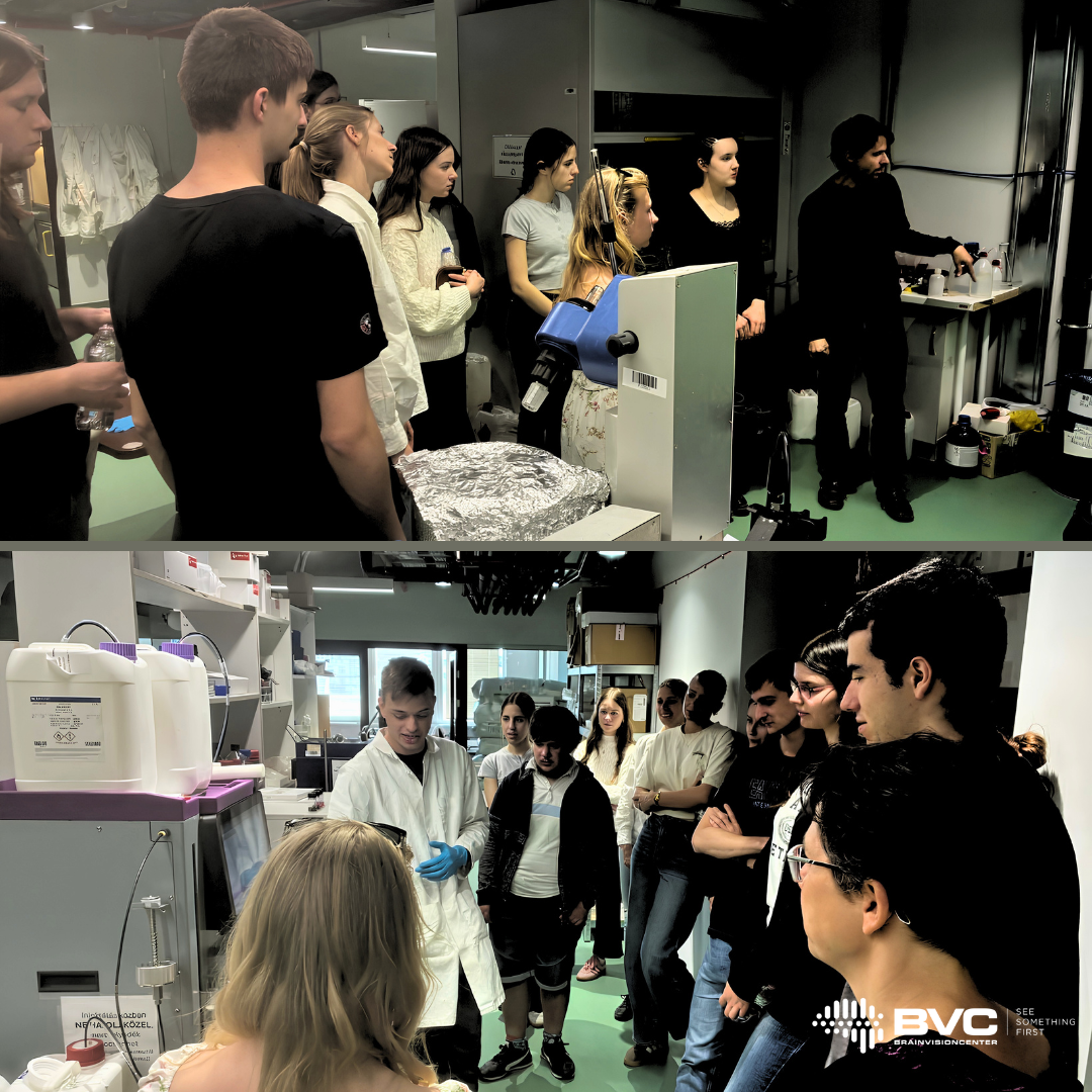
Beyond theoretical training, the program provides students with first-hand insight into some of Hungary’s leading research institutes. As part of this initiative, our institute recently welcomed a group of highly motivated students with a strong interest in chemistry and biology.
During their visit, our colleagues introduced them to our cutting-edge methodologies and state-of-the-art equipment-many of which represent world-class innovations in neuroscience research. The students gained a direct glimpse into how advanced scientific work is conducted in an internationally recognized research environment.
The experience proved to be highly inspiring, as reflected in their feedback:
“It was fantastic to see such instruments in operation.” “I was surprised by how many young researchers are working here with such dedication.” “I wouldn’t have thought that such high-quality work is carried out at a domestic research institute.”
We believe that encounters like these play a key role in motivating the next generation of scientists and fostering long-term interest in scientific careers.
A scientific paper by Dr. Balázs Rózsa (BrainVisionCenter, Institute of Experimental Medicine), Dr. Attila Losonczy (UT Southwestern Medical Center), and their co-authors was published in the March issue of Neuron, one of the world’s leading journals in neuroscience. The study, titled “Movement-stabilized three-dimensional optical recordings of membrane potential changes and calcium dynamics in hippocampal CA1 dendrites,” presents a three-dimensional real-time motion-correction method developed through the collaboration of BrainVisionCenter (BVC)—founded by Botond Roska and Balázs Rózsa—and Femtonics Ltd.
Figure 1. Real-time motion correction and single-cell dual-color labeling for motion-stabilized dendritic voltage-calcium imaging in vivo
This technology has, for the first time, made it possible to measure the brain’s elementary functional units—neurons and their processes, the so-called dendrites—in a continuously pulsating, moving brain, at the extremely fast timescales characteristic of brain activity, reaching speeds of several kilohertz. Under the leadership of Attila Losonczy and Balázs Rózsa, the researchers implemented rapid image stabilization together with simultaneous high-resolution, AI-assisted measurements in three dimensions.
The instrument—protected by more than 50 international patents—is manufactured and distributed by Femtonics Ltd. Its users include leading universities and research institutes such as Stanford University, Yale University, Columbia University, Harvard University, Boston University, the University of Oxford, McGill University, the University of Helsinki, MIT, and Caltech.
The publication in Neuron marks the 25th high-impact paper published in a Nature Index journal generated by the Institute of Experimental Medicine and BrainVisionCenter using a Femtonics instrument. This is considered a record achievement in the Hungarian high-tech sector. Globally, several hundred high-ranking scientific publications have been published using Femtonics laser microscopes. Revenue generated from the sale of this instrument platform has exceeded HUF 10 billion, which the company has reinvested in full into medical research and innovation.
The technology presented in the Neuron paper could enable intraoperative measurement of activity in the pulsating human brain, opening new avenues in medical diagnostics and therapy. It is capable of tracking motion of up to 1 mm while measuring the moving brain with 100-nanometer precision—approximately one-thousandth of the thickness of a human hair. Researchers at BVC and Femtonics are now working on the development of suitable sensors for human brain measurements, paving the way toward clinical application.
The key scientific breakthrough enabled by this invention is that dendritic integration and back-propagating action potentials—previously studied mainly in vitro—can now also be observed in awake, behaving animals, and tracked along the complex, tree-like branching processes of neurons, while filtering out motion-related artifacts arising from brain tissue movement, respiration, and heartbeat. The system can reposition the measurement coordinate system in less than 100 microseconds (i.e. 0.0001 seconds), thereby compensating for tissue displacement. In essence, it functions as a high-speed 3D image stabilizer used during laser scanning, resulting in stable images and signals free from motion noise.
One of the method’s major innovations is that, instead of using previously introduced artificial fluorescent beads in the brain, the neuronal soma itself can serve as the reference point for motion tracking. Because this approach does not trigger an immune response, it avoids damage to the brain and enables more accurate measurements. In addition, the method reduces motion-induced measurement artifacts by more than two orders of magnitude.
Figure 2. Electrical coupling properties between soma and dendrites across the dendritic arbor of hippocampal CA1 pyramidal neurons
Imaging based on the widely used calcium sensors provides only indirect information about electrical activity, and therefore offers a more temporally blurred view of brain function. By contrast, in the present study conducted by UT Southwestern Medical Center, BrainVisionCenter, and the Institute of Experimental Medicine, newly developed genetically encoded voltage sensors, used alongside calcium sensors, directly report electrical changes in the brain with a temporal precision of up to one ten-thousandth of a second. Until now, such measurements had not been feasible in vivo, at subcellular resolution and in 3D, because no sufficiently fast measurement technology existed.
The required technological breakthrough was provided by Femtonics’ acousto-optic technology, which enabled 3D measurements that are 6–7 orders of magnitude faster. This dual functional labeling approach allowed researchers to visualize electrical signals and the calcium responses they evoke at the same time. For labeling, Attila Losonczy’s group developed a novel rapid genetic delivery method based on in vivo electroporation, which yielded stronger contrast and better functional signals than any previous method, while leaving the brain unharmed.
The results show that responses measured in individual dendritic spines become progressively weaker as signals back-propagate along neuronal processes, the so-called dendrites. In contrast, trains of action potentials generate more sustained depolarization, allowing information to reach distal dendrites more reliably. Signals evoked by targeted photostimulation showed the same distance-dependent attenuation, suggesting that dendritic filtering arises primarily from the neurons’ own intrinsic properties. In addition, the researchers detected local dendritic voltage events that were independent of activity in the neuronal soma. This indicates that individual dendritic branches may function as partially autonomous computational subunits, almost like independent processors.
Another important finding is that—contrary to the currently prevailing view—the relationship between voltage signals and calcium signals weakens progressively as the number of neuronal branch points increases. In distal dendritic compartments, voltage signals remain measurable, but the corresponding calcium responses decline more rapidly, especially beyond branch points. This suggests that the structure and electrical properties of dendrites influence not only the propagation of voltage, but also calcium influx, thereby enabling branch-specific plasticity and molecular function.
These findings bridge the gap between in vitro dendritic measurements and in vivo neuronal dynamics. They establish a technological foundation for the direct study of dendritic computation during learning and memory formation in awake, behaving animals.
Nóra Lenkey and Máté Neubrandt, a young researcher couple who began their scientific careers at the Institute of Experimental Medicine, returned to Hungary after seven years at the University of Oslo to join BrainVisionCenter. Their work focuses on understanding how mice navigate and learn in both familiar and entirely new environments. Using a virtual reality system, mice run on a foam wheel while moving through colorful, computer-generated corridors displayed on monitors. This setup allows researchers to observe how internal spatial maps form in the hippocampus and how specific neuronal cell types influence an animal’s ability to orient itself, discover rewards, and adapt to novel surroundings. Their findings highlight the essential role of these cells in spatial navigation, suggesting parallels in human brain function.
BrainVisionCenter provides a unique environment where microscopy development, biological experiments, molecular tools, and chemical innovations are integrated, enabling rapid transitions from new concepts to practical applications. One of the center’s newest tools is a robot-arm–based microscope capable of following a freely moving mouse in real time, making it possible to perform high-resolution imaging during natural behavior and expanding the range of behaviors that can be studied under realistic conditions.
Nóra joined the epilepsy research group, which is developing a diagnostic method to precisely locate seizure-generating brain regions during surgery by imaging neuronal activity at the level of individual cells. This approach may enable surgeons to identify the exact cells involved in epileptic activity and potentially eliminate them with focused laser pulses, minimizing tissue loss and preserving critical brain functions. The method has already been validated in mice, and its publication is underway.
Their decision to return home was influenced by the scientific vision of the center, the opportunity to collaborate closely with engineers and developers, and the dynamic, translational research environment. On a personal level, they valued being closer to family, especially for their two young daughters, and found it difficult to fully integrate socially in Norway due to cultural distance. In Budapest, they appreciate the ease of forming connections and the familiar sense of community.