BrainVisionCenter’s international scientific network is shaped not only by high-impact publications, but also by active collaborations with leading researchers from around the world.
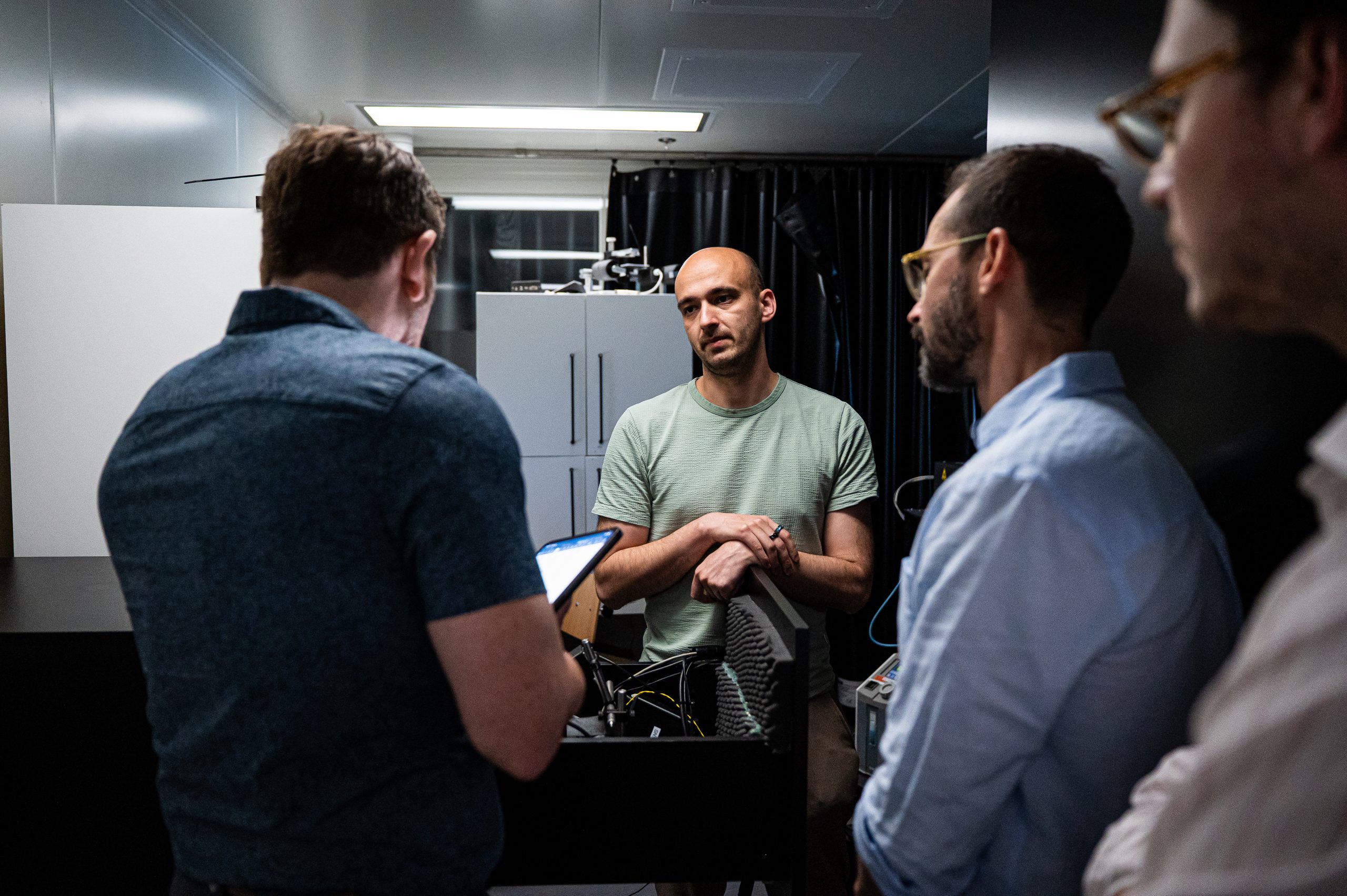
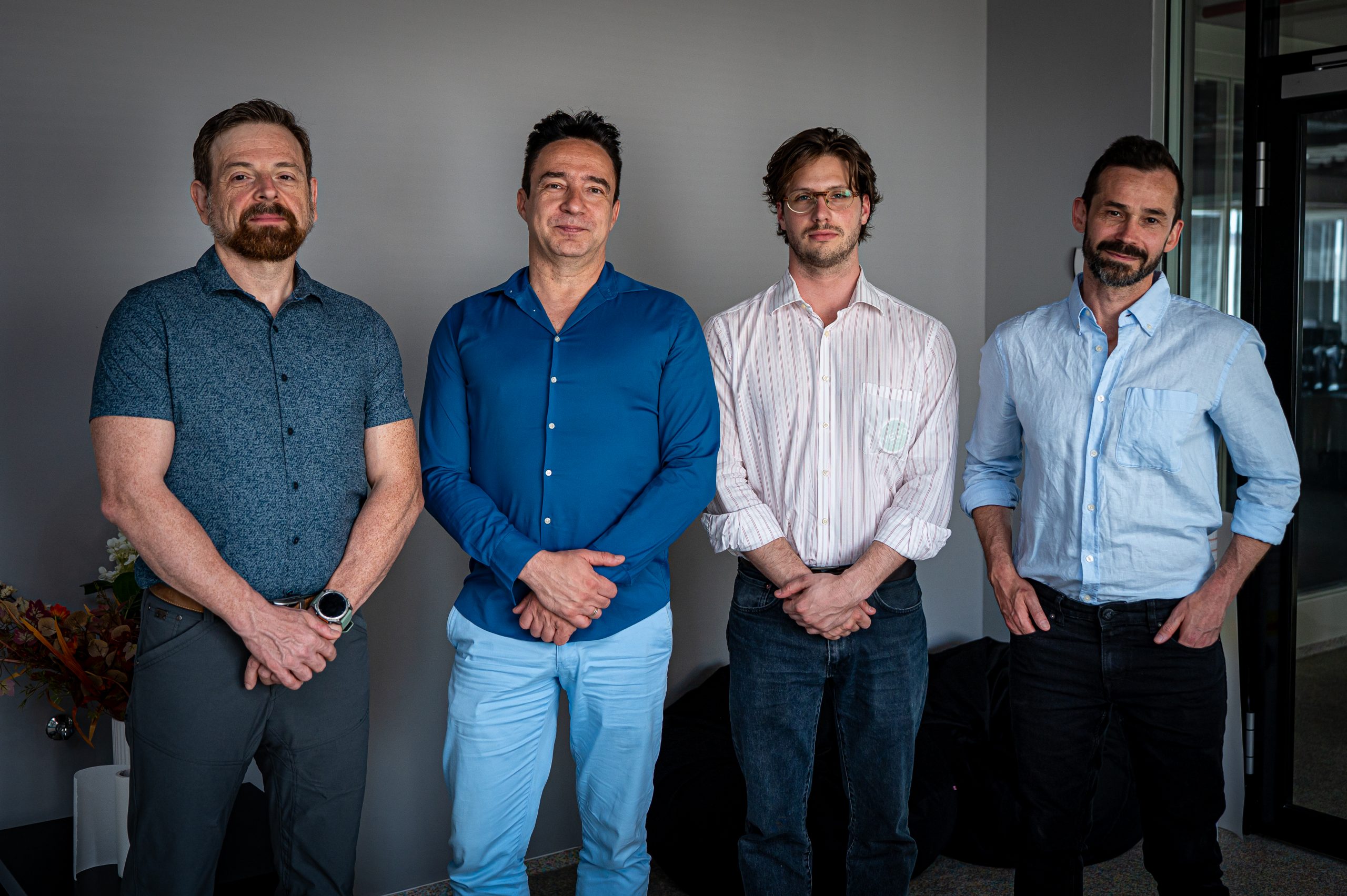
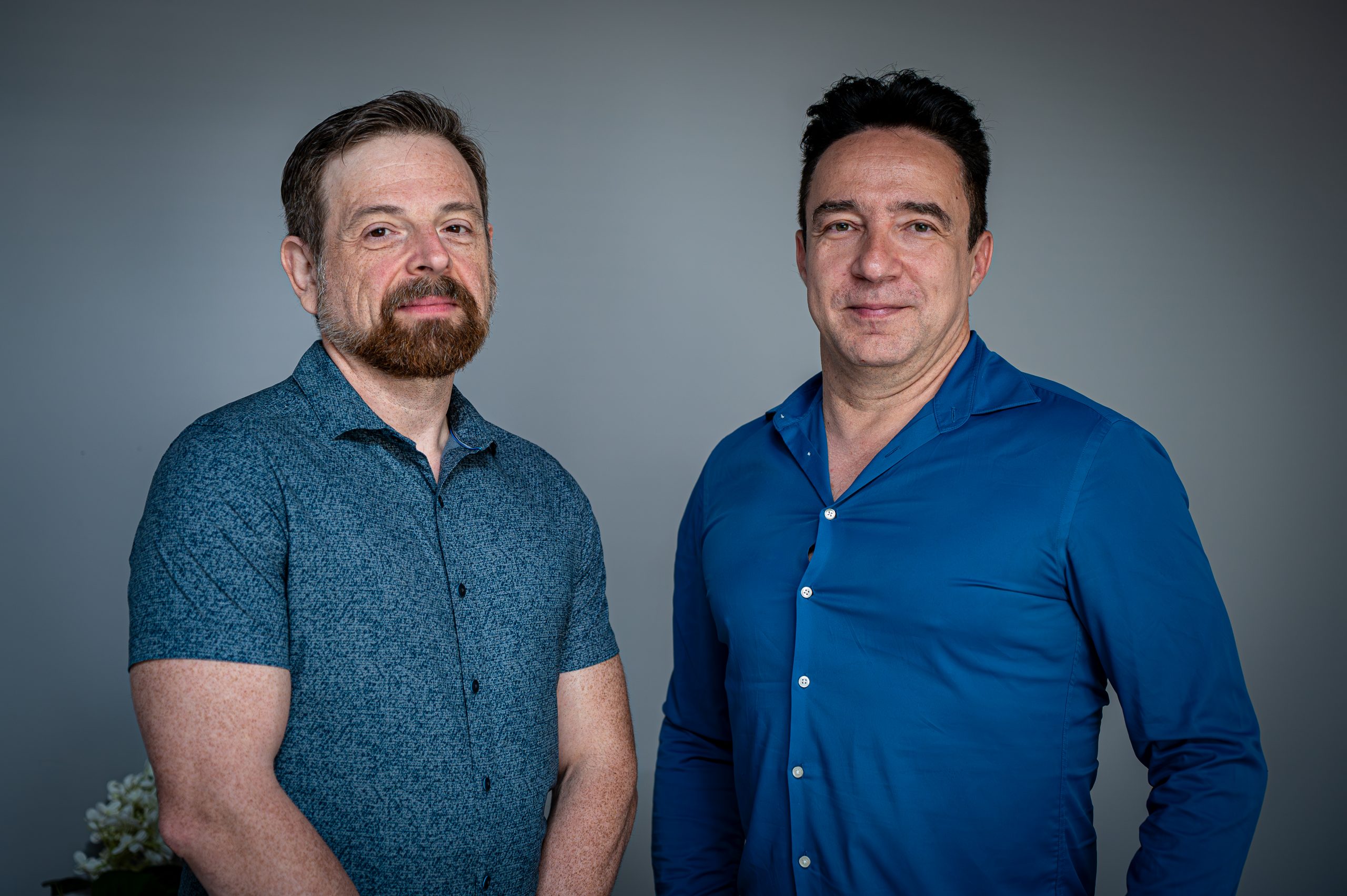
Recently, Professor Attila Losonczy, a leading neuroscientist at UT Southwestern Medical Center, visited our institute to discuss ongoing and future joint research directions. Professor Losonczy and our founder, Dr. Balázs Rózsa, have been connected through several scientific collaborations combining advanced neuroscience, cutting-edge optical imaging, and innovative experimental approaches.
This partnership has already contributed to significant scientific results, including a recent joint publication in Neuron, one of the world’s leading neuroscience journals.
During the visit, the teams reviewed current research activities and defined several new collaborative projects. These future directions mark the next chapter in a long-standing scientific partnership and further strengthen BrainVisionCenter’s connection to the international forefront of neuroscience research.
Through collaborations like this, BrainVisionCenter continues to build bridges between advanced imaging technology, experimental neuroscience, and world-class scientific expertise.